The QRS complexes show an abnormal morphology with a width greater than the baseline QRS complex often 011 second. Giemsa stain also is used to stain Histoplasma capsulatum Pneumocystis jiroveci Klebsiella granulomatis Talaromyces marneffei formerly called Penicillium marneffei and occasionally bacterial capsules.

Histopathologic Diagnosis Of Fungal Infections In The 21st Century

Histoplasma Capsulatum Fungal Infections Antiinfectivemeds Com
Bone Marrow Aspirate Showing Histoplasma Capsulatum
Bronchoalveolar or peritoneal macrophages for Histoplasma capsulatum.

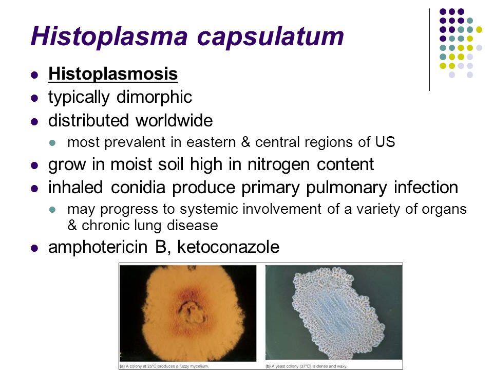
Histoplasma capsulatum morphology. Of the six systemic agents five Histoplasma capsulatum Blastomyces dermatitidis Paracoccidioides brasiliensis Coccidioides immitis and Penicillium marneffei are dimorphic changing from a mycelial to a unicellular morphology when they invade tissues except C immitis that forms spherules. Morphology and General Properties of Fungi MICROBIOLOGY MODULE Microbiology Notes Coccidioidomycosis Self-limited respiratory disease Blastomyces dermatltidis orchronic progresssive infection of various organs Histoplasmosis Fungus infection of the lungs Coccidioides immitis with fever. Occasionally other organs are affected.
The filamentous hyphal and single celled budding forms yeast. Diagnosis of histoplasmosis by antigen detection based upon experience at the histoplasmosis reference laboratory. The opportunistic fungal pathogens include Cryptococcus neoformans Candida Aspergillusspp Penicillium marneffei the Zygomycetes Trichosporon beigelii and Fusarium spp.
The primary systemic fungal pathogens include Coccidioides immitis Histoplasma capsulatum Blastomyces dermatitidis and Paracoccidioides brasiliensis. Histoplasma primarily infects lungs but can spread to other tissues causing histoplasmosis a potentially fatal disease. But often we can get diagnostic clues just by looking at the morphology of the RBCs on a smear and linking their appearance to the most likely causes.
Mediastinal lymphadenopathy is a feature of primary TB and is a common finding on chest radiographs of children with TB disease. More than normal RBCs. Program within mayoclinicgradschool is currently accepting applications.
Symptoms of this infection vary greatly but the disease affects primarily the lungs. It is associated with a variety of dermatological conditions caused by fungal infections notably seborrhoeic dermatitis and tinea versicolorAs an opportunistic pathogen it has further been. Human pathogenic fungi produce three basic cell types.
Hyphae yeast cells and spores. RBC Morphology 300 RBC Inclusions 303 Staining of RBC Inclusions 305 Erythrocyte Indices 306 Hemoglobinopathy versus Thalassemia 307 Normocytic Anemias 308 Macrocytic Anemias 310 Microcytic Hypochromic Anemias 311 Differentiation of Microcytic Hypochromic Anemias 312 Acute versus Chronic Blood Loss 313 Granulocytic Maturation 314. Growth of fastidious pathogenic fungi such as Histoplasma capsulatum and Blastomyces dermatitidis.
22 23 In contrast postprimary TB of HIV-seronegative adults is characterized by cavitation and poorly defined consolidation of the apical and posterior. Cross-reactivity in Histoplasma capsulatum variety capsulatum antigen assays of urine samples from patients with endemic mycoses. Histoplasma primarily infects lungs but can spread to other tissues causing histoplasmosis a potentially fatal disease.
The organization and subcellular structure of these different cell types and their modes of growth and formation are reviewed. Cornmeal agar Cornmeal tween agar. Zhou P Sieve MC Bennett J Kwon-Chung KJ Tewari RP et al.
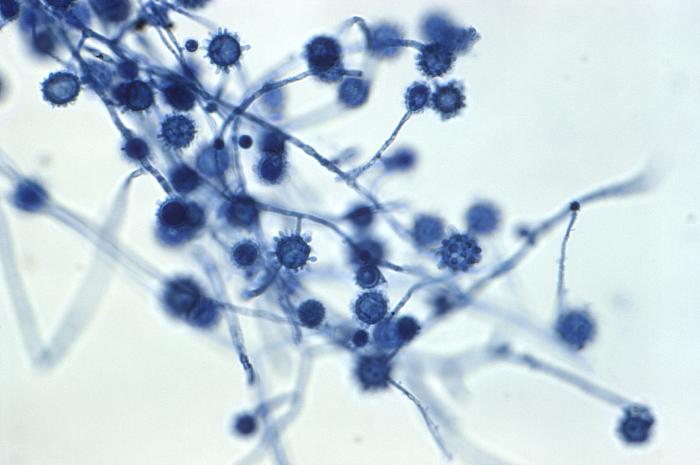
1995 IL-12 Exp Immunol 102. The dark cells in this bright field light micrograph are the pathogenic yeast Histoplasma capsulatum seen against a backdrop of light blue tissue. Curr Opin Microbiol 3.
Helmuth Reuter Robin Wood in Tuberculosis 2009. Histoplasma capsulatum Blastomyces dermatidis Paracoccidiodes brasiliensis Coccidioides immitis Some 200 human pathogens have been recognized from among an estimated 15 million species of fungi. 48 Likes 2 Comments - College of Medicine Science mayocliniccollege on Instagram.
Indeed the incubation time required to acquire fungal growth is one diagnostic indicator used to identify or confirm fungal species. Histoplasmosis is a chronic respiratory infection caused by inhaling the spores of the fungus Histoplasma capsulatum which is found in. Clin Infect Dis 24.
The incidence of these infections is increasinglargely because of rising numbers of immunocompromised patients including those with neutropenia human immunodeficiency virus chronic immunosuppression indwelling prostheses burns and diabetes. Tuberculate macroconidia were seen on the hyphae of the mold phase. Histoplasmosis is a fungal infection caused by Histoplasma capsulatum.
Incubation times will vary from approximately 2 days for the growth of yeast colonies such as Malasezzia to 2 to 4 weeks for growth of dermatophytes or dimorphic fungi such as Histoplasma capsulatum. Peptone glucose chloramphenicol Chromogenic ix agar. Prevents mortality in mice infected with Histoplasma capsulatum through 22.
Loss of Histoplasma capsulatum weightenlargement. Ehrlichia canis Histoplasma capsulatum Dipetalonema reconditum and Dirofilaria immitis heartworm. Called disseminated histoplasmosis it can be fatal if left untreated.
The dark cells in this bright field light micrograph show budding in the pathogenic yeast Histoplasma capsulatum seen against a backdrop of light blue tissue. Malassezia furfur formerly known as Pityrosporum ovale in its hyphal form is a species of yeast a type of fungus that is naturally found on the skin surfaces of humans and some other mammals. Growth and form is the consequence of how new cell surface is formed.
A fungal isolate from the sputum of a patient with a pulmonary infection is suspected to be Histoplasma capsulatum. This is generated by the delivery of vesicles to the surface which provides new membrane and the enzymes. Granulomatous inflammation is a distinctive form of chronic inflammation produced in response to various infectious autoimmune toxic allergic and neoplastic conditions It is defined by the presence of mononuclear leukocytes specifically histiocytes macrophages which respond to various chemical mediators of cell injury.
Fungi exist in two fundamental forms. This stain is also used in cytogenetics to stain the. 11691171 Google Scholar Wheat LJ Garringer T Brizendine E Connolly P 2002.
The colony morphology and microscopic characteristics are consistant with M audounii. Selective and differential chromogenic medium for the isolation and identification of various Candida species. Invasive fungal and fungal-like infections contribute to substantial morbidity and mortality in immunocompromised individuals.

Histoplasma Introduction Morphology Life Cycle Pathogenesis Labora
Microscopic Morphologic Features Of The Mold Forms Of Various Dimorphic Fungi Medical Laboratories

Disseminated Histoplasmosis In Immunocompetent Patients From An Arid Zone In Western India A Case Series Sciencedirect

Fungal Pathogens Shape Shifting Invaders Trends In Microbiology

Histoplasma Capsulatum In Peripheral Blood Smear Bmj Case Reports

Histoplasmosis Wikipedia

Details Public Health Image Library Phil

Histoplasma Capsulatum Microbewiki

